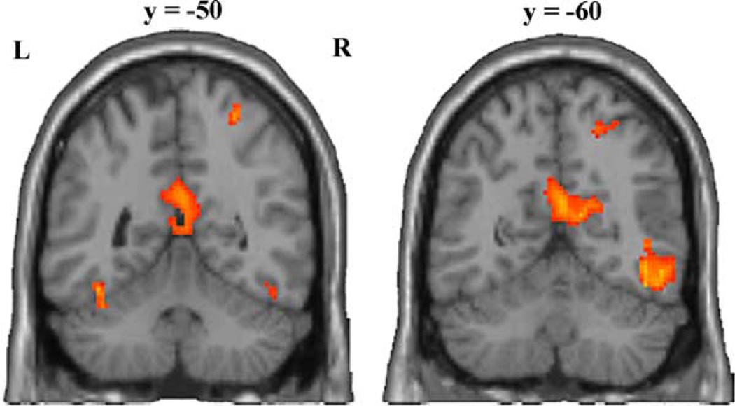
Fig. 3.
Two coronal sections of a representative brain showing activity associated with imitating. The anterior section (left) shows clusters of activity in the posterior cingulate, extending to the rostral end of the calcarin sulcus and bilaterally in the fusiform gyrus. The most posterior section shows clusters of activity in the calcarin sulcus and extrastriate body area in the right hemisphere. Both sections show activity in the right intraparietal sulcus.

