
Figure 3.
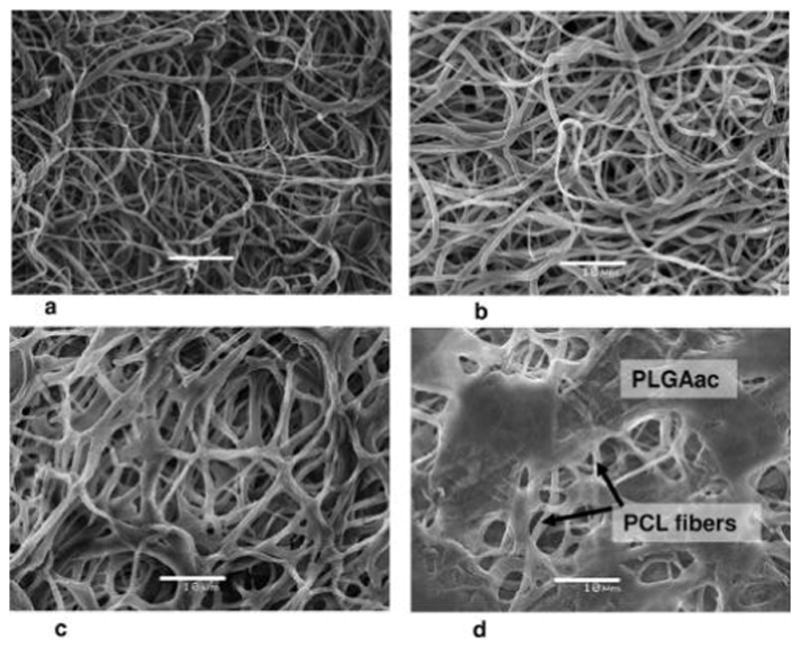
Representative SEM micrographs of the PCL-PLGAac nanofibrous scaffold with an internal compositional gradient. Images were taken from the PLGA side. (a) Morphology of as spun PLGAac nanofibers. (b–d) Changes in the nanofiber morphology after 1 week (b), 3 weeks (c), and 5 weeks (d) of degradation in phosphate buffer solution at 37 °C. The unchanged PCL nanofibers underneath the PLGAac layer were found after 5 weeks of degradation (d). Scale bar shows 10 μm.