Figure 1.

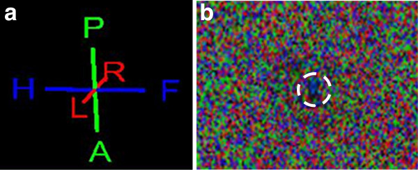
a. Color-coding of the directional fiber tracking according to the course of water diffusion. The data were color coded with blue indicating head-to-feet or craniocaudal direction, red denoting right-to-left (R-L) direction, and green indicating anterior-to-posterior (A-P) or dorsoventral orientation of water diffusion. b. DTI raw data image with water proton diffusion coloring (as described above). In the center (white circle) a relatively large blue colored area is visible caused by the craniocaudal course of water diffusion which is used to identify the spinal cord.
