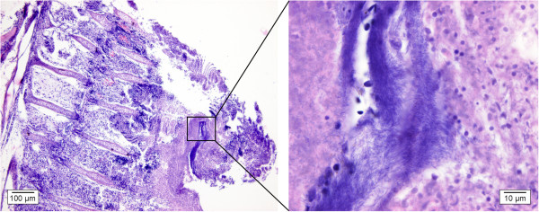
Figure 6.

Gill section of a koi carp infested with F. columnare. In the left figure, extensive loss of branchial structures is visible. This is an advanced stage of the disease in which the filaments and the lamellae have fused and the gill epithelium is destroyed (H&E, bar = 100 μm). Complete clubbing of gill filaments may finally result in circulatory failure and extensive internal hemorrhage. A detail of this is depicted in the right figure, where F. columnare bacteria are visible as long, slender, purplish structures in between remnants of the gill tissue (H&E, bar = 10 μm).
