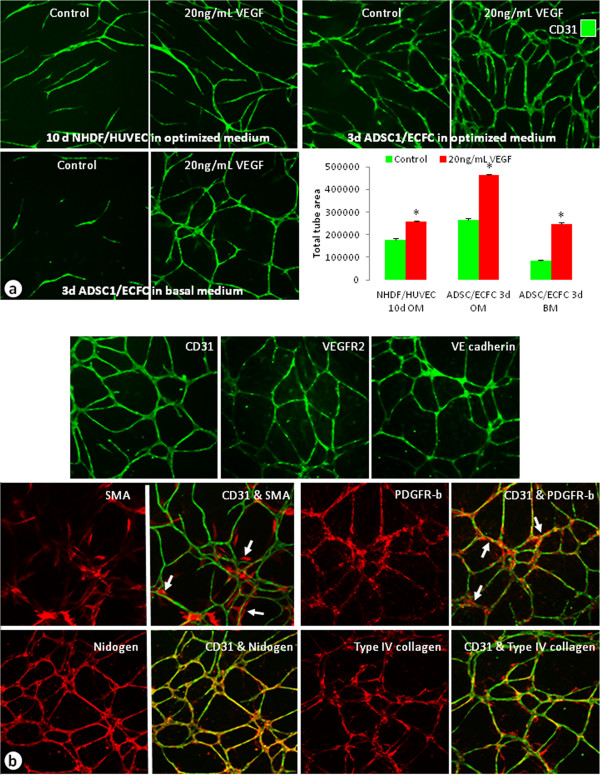
Figure 1.
Characterization of co-cultured cord formation assays. (a) Unstimulated or VEGF-stimulated (20 ng/mL) cords stained with CD31 from co-cultures of NHDFs and HUVECs (top left), ADSCs and ECFCs in optimized medium (top right), and ADSCs and ECFCs grown in basal medium (bottom left). Graph compared the total tube areas of the cords from the different assay systems. n = 3–5 per group. * = p < 0.0001 vs. respective control. (b) Images of 5d ADSC and ECFC cords grown in basal medium and stimulated with 20 ng/mL VEGF. Endothelial cells were labeled with CD31, VEGFR-2, or VE-cadherin (top), mural cells or pericytes were labeled with SMA or PDGFR-β (middle), and vascular basement membrane was identified by nidogen and type IV collagen antibodies (bottom). Arrows indicate areas where pericytes labeled with SMA or PDGFR-β were associated with the cords.

