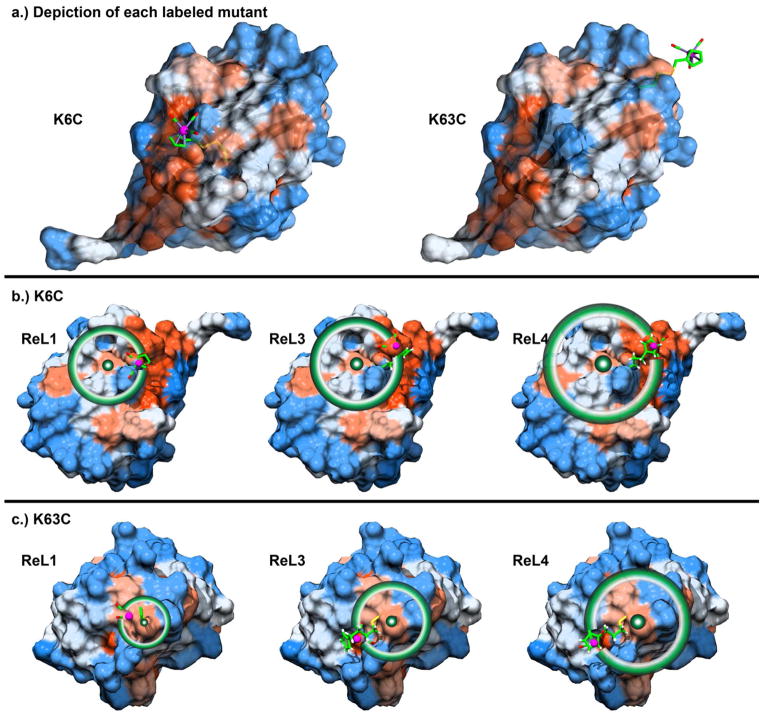
Figure 4.
Hydrophobicity of the ubiquitin surface. (a) Side view of the two labeled positions of ubiquitin, K6C and K63C with ReL1 label attached. Schematics of distances accessible to ReL1, ReL3, and ReL4 labels attached to (b) K6C and (c) K63C with circles drawn about the cysteine β-carbon. Hydrophobic and hydrophilic residues are shown red and blue, respectively, according to the Kyte-Doolittle hydrophobicity scale, as described in the SI.

