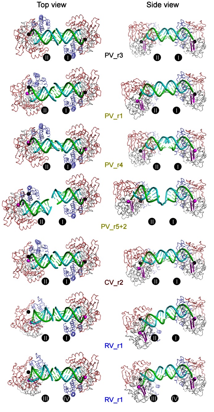
Figure 3. A comparison of inter-complex RNA-RNA junctions.
Cartoon representations of seven EC pairs depicting the variety of inter-complex junctions observed in the crystals. The right side polymerase within each pair is always shown in the same orientation. The polymerases and RNA follow the color-coding scheme from Figure 1 with the YGDD residues of conserved motif C colored in magenta to help locate the polymerase active site. The PV_r3 structure (top) is used as a reference with its active site Asp328 Cα atoms shown as black spheres. These two black spheres are then overlaid onto the other complex pairs where the Cα atoms of the corresponding Asp residues are shown in magenta to highlight the differences in the placement of the non-superimposed left-side ECs. Non-crystallographic symmetry relationships show that all ECs except RV_r1 obey a pseudo two-fold symmetry within each dumbbell pair. See Figure 4 for a superpositioning of all the complexes.

