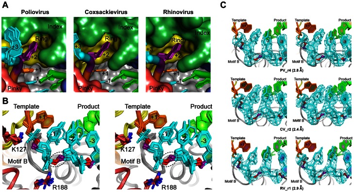
Figure 7. Conservation of RNA active site conformation.
A) Top view looking at the downstream template RNAs from all ECs as it enters the active site. The +2 template strand nucleotide (purple) is consistently bound in a pocket formed by the polymerase index and ring fingers and the templating +1 nucleotide (orange) is always fully pre-positioned for base pairing with an incoming NTP in the active site. B) Stereo images of the RNA template strand conformation from positions −3 to +1 showing RdRP motif B positioned directly beneath the -1 position ribose of the template strand. The backbone of the -2 template strand nucleotide always has a different conformation than the neighboring A-form RNA helix nucleotides that is due to a 120° rotation about its ribose C5′-O5′ bond (dashed circle). C) Stereo images of composite SA-omit electron density maps (3500 K, 1.5 σ) showing RNA density in the vicinity of the conserved non-standard backbone conformation of the −2 template strand nucleotide.

