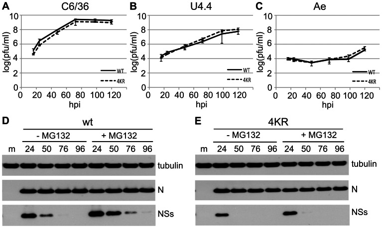
Figure 6. wtBUNV and rBUN4KR infection of mosquito cells.
(A–C) Virus growth in mosquito cells. Triplicate wells of C6/36, U4.4, or Ae cells were infected at 0.01 PFU/cell and supernatants analysed by plaque assay titration. (D, E) Expression of NSs protein. C6/36 cells were infected with wtBUNV (D) or rBUN4KR (E) at 5 PFU/cell, and left untreated or treated with MG132 for 8 h prior to harvesting. Cell lysates were analysed by immunoblotting using antibodies against the proteins indicated to the right. Above the lanes, m = mock; numbers are hpi.

