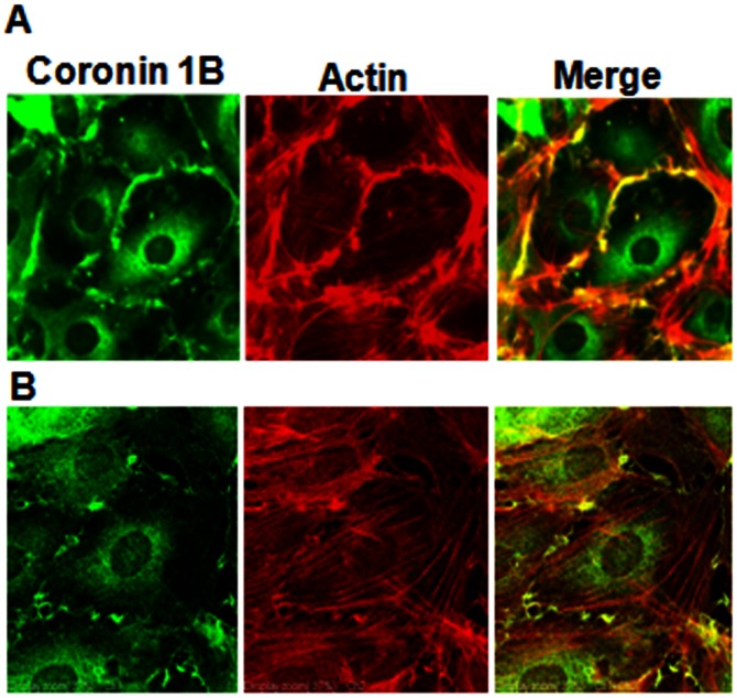
Figure 2. Coronin 1B localization in human lung endothelial cells.
HPAECs grown to ∼90% confluence on slide chambers were fixed, permeabilized and localization of Coronin 1B, actin and co-localization of Coronin 1B with actin was visualized by immunocytochemistry as described in Materials and Methods. Shown are representative immunofluorescence images from several independent experiments as measured by regular (A) immunofluorescence and (B) confocal microscopy.

