
Figure 3. Effect of starvation and serum supplementation on Coronin 1B localization in human lung endothelial cells.
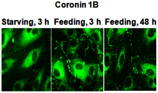
HPAECs grown on slide chambers (∼90% confluence) were incubated in EBM-2 medium containing either 0.1% serum for 3 h or in EBM-2 medium containing 5% serum for 3 h and 48 h. Cells were fixed, permeabilized and Coronin 1B localization was visualized by immunocytochemistry as described in Materials and Methods. Shown are representative immunofluorescence images from several independent experiments.