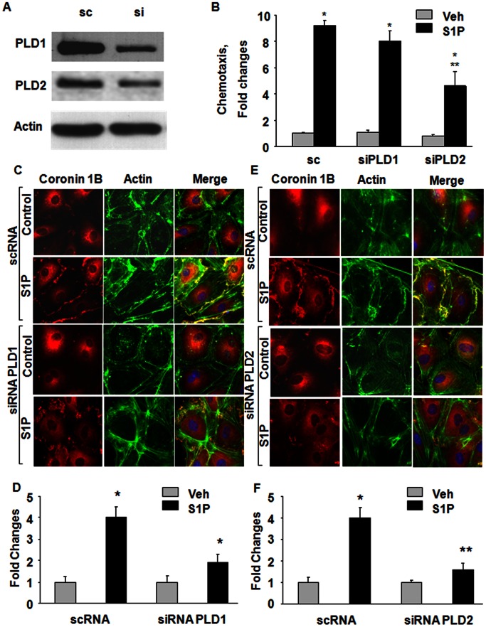
Figure 6. Role of PLD2 in S1P-induced chemotaxis, Coronin 1B and actin lamellipodia localization in HPAECs.
HPAECs (∼50% confluence) were transfected with scrambled (sc), PLD1 or PLD2 siRNA (50 ng/ml) for 72 h. (A) Cell lysates (20–40 µg of protein) were subjected to 10% SDS-PAGE, Western blotted and probed with PLD1 and PLD2 antibodies as indicated; (B) chemotaxis of scrambled (sc) or siRNA transfected cells to S1P (1 µM) for 15 min was carried out in a Boyden chamber-based trans-well assay as described under Materials and Methods. Values are mean±SEM of three independent experiments in triplicate. *, p<0.01 compared cells without S1P; **, p<0.005 compared to scrambled siRNA transfected cells plus S1P; HPAECs transfected with sc, PLD1 (C) or PLD2 (E) siRNA in 100-mm dishes as described under (A) were trypsinazied and seeded onto slide chambers prior to stimulation with S1P (1 µM) for 15 min. Cells were washed, fixed, permeabilized, and probed with anti-Coronin 1B and AlexaFluor Phalloidin antibodies, and redistribution of Coronin 1B and actin due to downregulation of PLD1 (D) or PLD2 (F) was examined by immunofluorescence microscopy using a 60 X oil objective and quantified by ImageJ software as described under “Experimental Procedures”. Shown is an immunofluorescence micrograph from three independent experiments.

