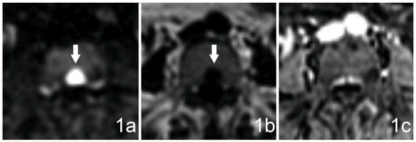
Figure 1.

Examples of the conspicuity scale in a 53 year old woman with breast cancer. (a) A bone lesion in the posterior aspect of the L4 vertebral body demonstrates restricted diffusion, is highly conspicuous (arrows), and has a score of 5 on the five-point conspicuity scale. (b) The same lesion is slightly less conspicuous on the FO image generated during the same T1-weighted fast Dixon-based acquisition and has a score of 3 on the conspicuity scale. (c) The lesion is not visible on the FS T1-weighted + C image and thus has a score of 1 on the conspicuity scale. The bone lesions in our study exhibited variable conspicuity on different sequences, and the same patient often had multiple lesions with widely varying conspicuity patterns. The differing lesion conspicuity on the various sequences exemplified the need for obtaining multiple sequences in order to increase the likelihood of effectively detecting bone metastases.
