
Figure 2.
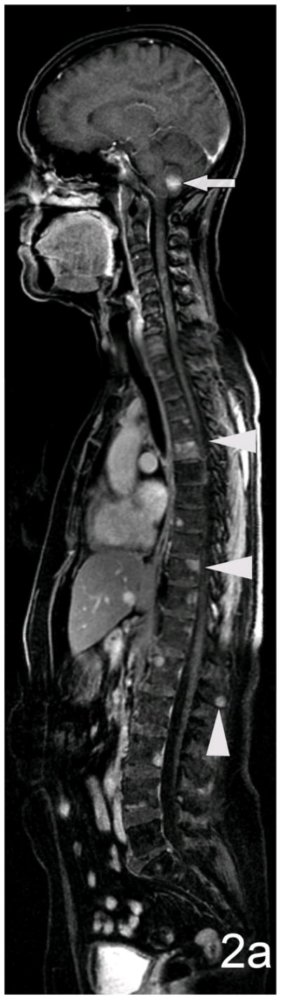
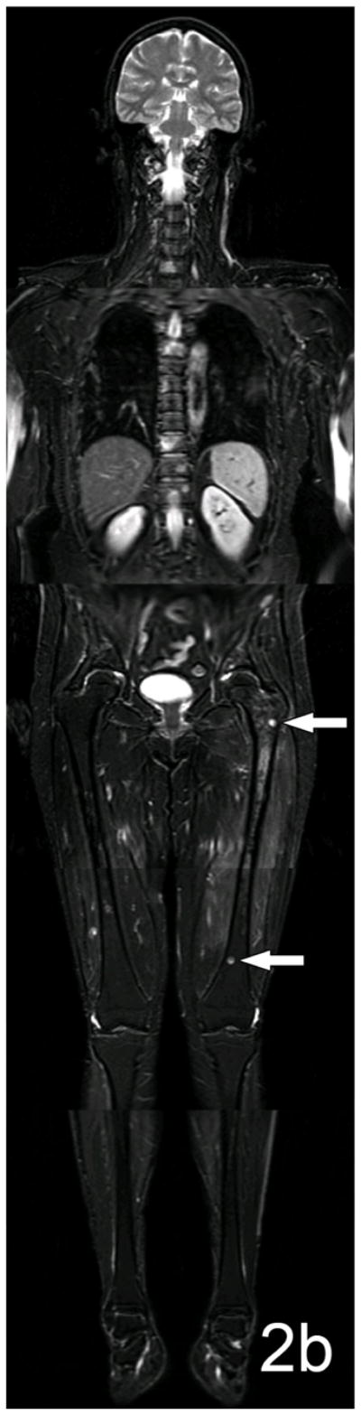
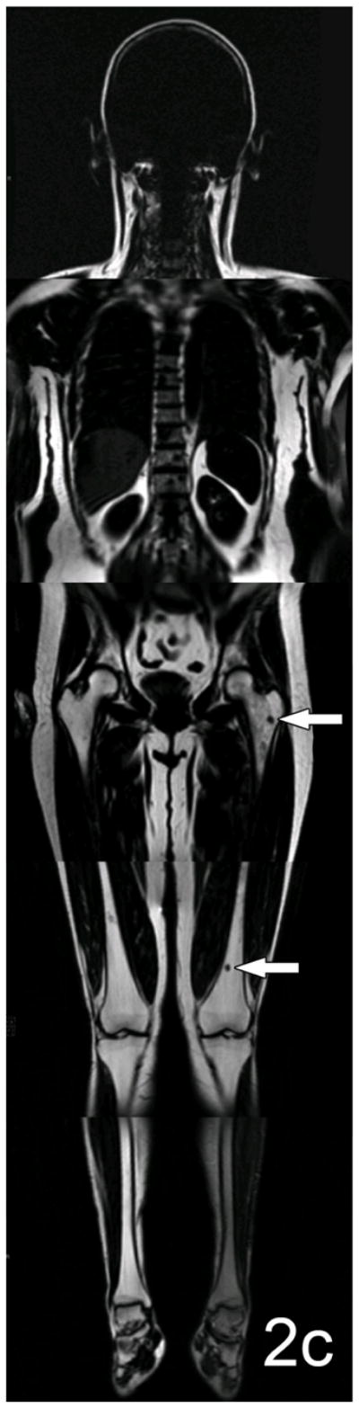
Sagittal and coronal composite images demonstrating whole-body lesion conspicuity. Figure (a), is a sagittal composite of FS T1 + C images in this 55 year old woman with breast cancer and is the sequence upon which bone metastases were most conspicuous in the study. Enhancing metastases in numerous vertebral bodies are readily apparent on a background of darkly saturated fat. Three of the most conspicuous bone metastases are indicated (arrowheads) and rank 4–5 on the conspicuity scale. An enhancing brain metastasis is incidentally detected (arrow). On the coronal composite images (b, c) focal metastases in the spine and proximal and distal left femur are shown (arrows). The lesions on the coronal FS T2-weighted image (b) are bright on a background of saturated fat. The lesion in the proximal left femur (upper arrow) enhances well, is highly conspicuous and ranked a score of 5 on the conspicuity scale. The lesion in the distal femur (lower arrow) is less conspicuous and ranks a 4. The lesions on the coronal FO sequences generated during the same acquisition (c) are dark on a background of bright fat. The lesion in the proximal femur is now ranked a 4 on the conspicuity scale because adjacent red marrow is also dark on FO images and the overall effect is to decrease the conspicuity of the lesion. Nevertheless, the lesion in the distal femur is surrounded by fatty yellow marrow and now is ranked 5 because of the high conspicuity produced between the dark lesion and the bright yellow marrow.