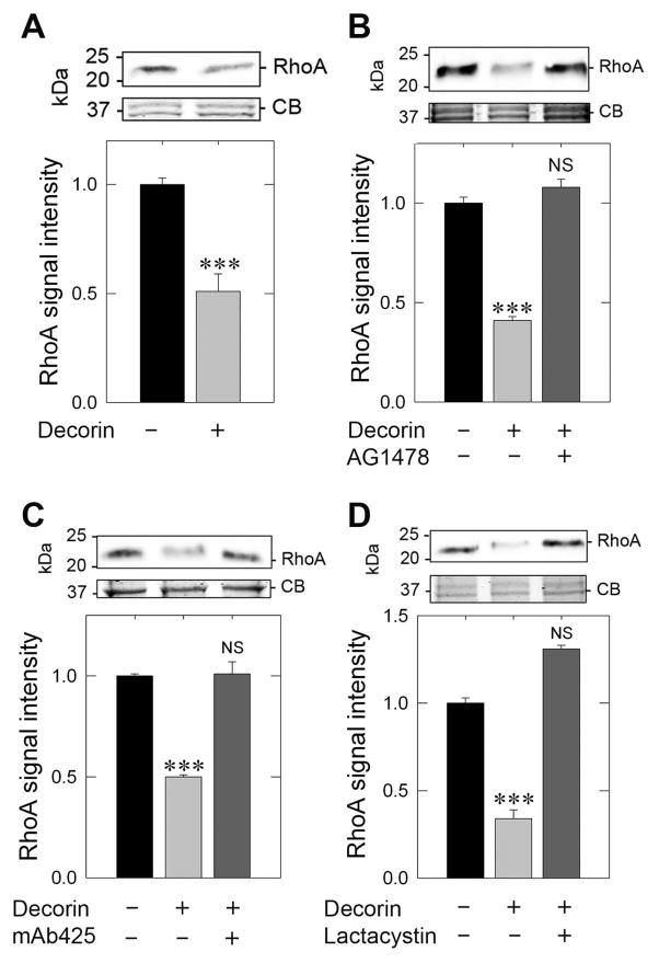
Fig. 4.
Decorin depends on EGFR to rapidly degrade RhoA and evoke rapid TSP-1 secretion. (A) Immunoblot analysis of RhoA in response to 200 nM decorin for 10 min in MDA-MB-231 cells (top panel) and quantification of RhoA levels (bottom panel). (B) Immunoblot analysis of RhoA after exposure to decorin alone (200 nM, 10 min) or following pretreatment (30 min) with the EGFR inhibitor, AG1478 (1 μM) (top panel), with accompanying quantification of RhoA levels (bottom panel). (C) RhoA analysis following decorin treatment (200 nM, 10 min) or after pretreatment (30 min) with the EGFR blocking antibody, mAb425 (10 μg·mL−1) (top panel) and corresponding quantification (bottom panel). (D) RhoA exposed to decorin alone (200 nM, 10 min) or in conjunction with a 30 min pretreatment of the proteasome inhibitor, lactacystin (10 μM) (top panel) and quantification (bottom panel). In all instances, the upper portion of the gel was Coomassie blue stained for equal loading and normalization of RhoA signal intensity. The data reported are representative of at least 3 independent experiments and expressed as the average fold change ±SEM. *** P < 0.001.

