
Fig. 5.
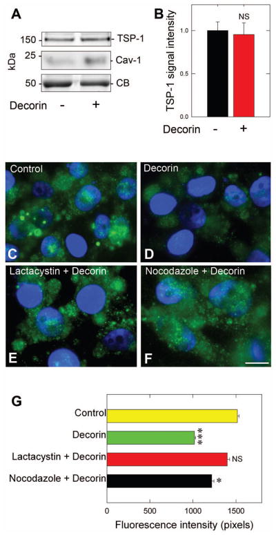
Decorin depends on classical secretory pathway for rapid TSP-1 secretion. (A) Immunoblot analysis of TSP-1 positive exosomes from MDA-MB-231 in response to 200 nM decorin (6 h) followed by ultracentrifugation (100,000 · g, 90 min) to isolate the exosome-containing fraction. (B) Quantification of exosomal TSP-1 after normalization to Coomassie blue. (C–F) Representative immunofluorescence images of MDA-MB-231 cells probe for TSP-1 (green) for control (C), decorin-only treated (200 nM, 10 min) or after pre-treatment (30 min) with the proteasome inhibitor, lactacystin (10 μM) as in (E) or with the microtubule inhibitor nocodazole (100 ng·mL−1) as in (F). The nuclei appear blue due to DAPI. All images were collected at the same exposure, gain, and intensity. (G) Quantification of average TSP-1 fluorescence intensity (n=10/treatment). Data are representative of at least three independent experiments and reported as normalized fold changes ±SEM (B), or as the average fluorescence intensity ±SEM for the reported immunofluorescence as in (G). * P < 0.05; ***P < 0.001; NS, not significant.