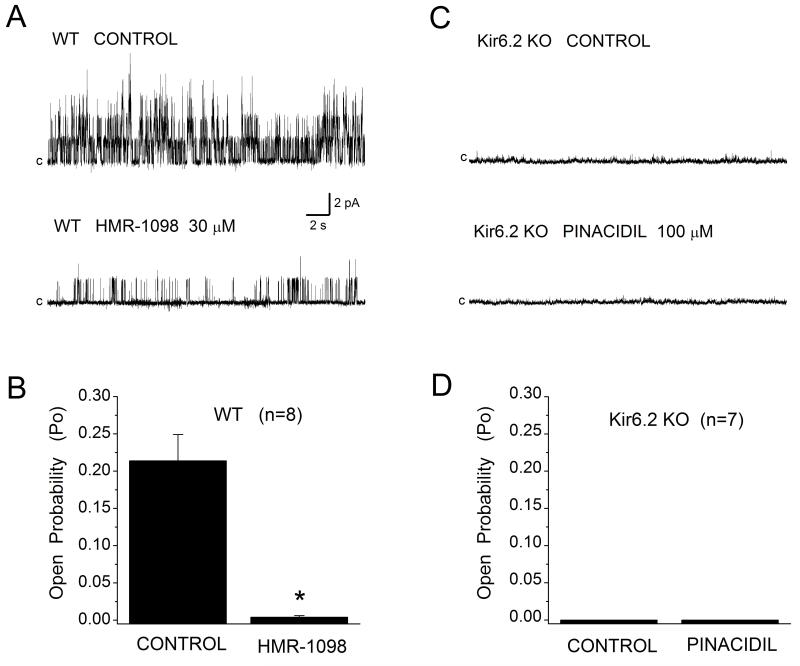
Figure 2.
The sarcKATP channel in mouse ventricular cardiomyocytes. (A) Single channel activity was monitored in the inside-out patch configuration at Em +40 mV in the absence (Control) or presence of sarcK channel blocker HMR-1098. (B) HMR-1098 (30 μM) inhibited channel activity by 97%. Data are mean±SEM, n=8. (C, D) No channel activity was detected in membrane patches excised from Kir6.2 KO myocytes when monitored in control conditions or during application of channel opener pinacidil (100 μM), n=7.

