Figure 4.
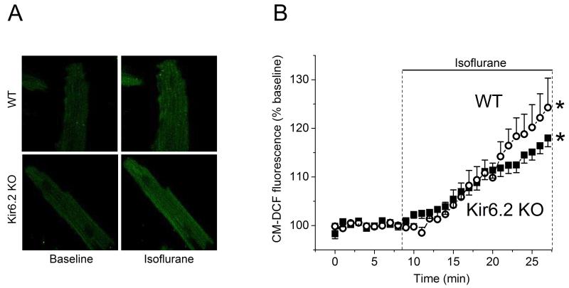
Acute isoflurane alters the mitochondrial redox state of intact mouse ventricular cardiomyocytes. Autofluorescence of mitochondrial flavoproteins (FP) was monitored as the indicator of mitochondrial redox state. (A) Example images of FP fluorescence captured in WT and Kir6.2 KO cardiomyocytes before (Baseline), during (Isoflurane) and after (Washout) exposure to 0.5 mM isoflurane. (B) Time course of changes in FP fluorescence during application of isoflurane. Data are expressed as change from the baseline (100%), data points are mean±SEM. Isoflurane increased oxidation of mitochondrial FP in cardiomyocytes from WT (closed symbols, n=6) and Kir6.2 KO (open symbols, n=5) hearts. *P< 0.05 vs. baseline. Washout between 14 and 25 min.

