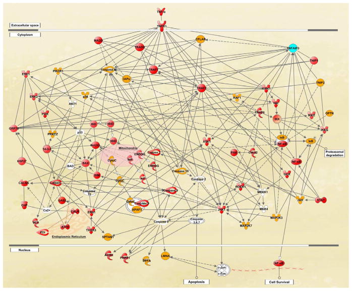
Figure 4.
Global proteomics analysis of the glaucomatous human retina. To determine alterations in retinal protein expression, retinal protein samples obtained from human donor eyes with or without glaucoma were analyzed by label-free quantitative 2D-LC-MS/MS analysis. The knowledge-based analysis of the generated high-throughput datasets established extended networks of diverse functional interactions between death-promoting and survival-promoting pathways and mediation of immune response. Up-regulated pathways included death receptor-mediated caspase cascade, mitochondrial dysfunction, endoplasmic reticulum stress, and calpains leading to apoptotic cell death; and NF-κB and JAK/STAT pathways, and inflammasome mediating inflammation. Proteins shown in red color exhibited significantly increased expression and shown in yellow color no significantly increased expression in glaucomatous samples relative to non-glaucomatous controls, while the protein shown in blue color exhibited prominent individual differences. Proteins shown in white color were not detectable by quantitative LC-MS/MS. This simplified network, generated using the Ingenuity Pathways Analysis, has recently been published (Yang et al., 2011).

