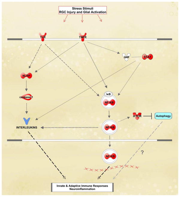
Figure 7.
I nflammatory responses of astrocytes in ocular hypertensive rat retinas. In this simplified network generated by The Ingenuity Pathways Analysis, astrocyte proteins shown in red color exhibited significantly increased expression in ocular hypertensive samples relative to normotensive controls. Based on the high-throughput proteomics data validated by Western blot analysis and immunohistocemical labeling, TNF-α/TNFR signaling, NF-κB activation, JAK/STAT signaling, TLR signaling, and inflammasome appear to be co-players of inflammatory responses mediated by ocular hypertensive astrocytes. This figure has recently been published (Tezel et al., 2012c).

