
Fig. 2. Increased phospho-ERK1/2-staining in cortical slices in Mg2+-free condition with GABAA-R blockade.
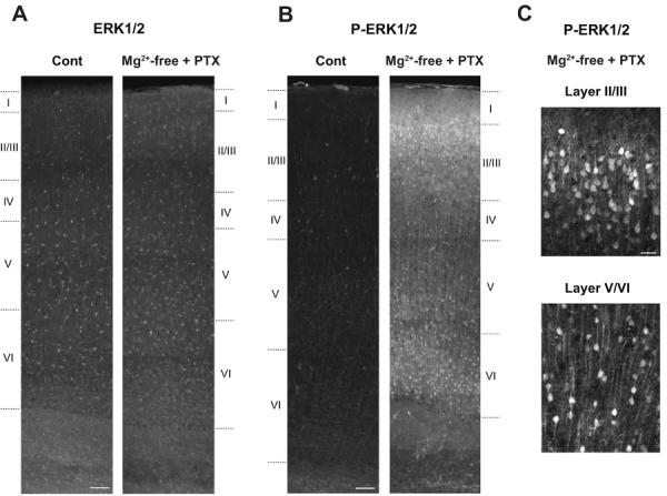
Representative staining with anti-ERK1/2 (A) and with anti-phospho-ERK1/2 (P-ERK1/2) (B) in control slices incubated in normal ACSF containing 1.2 mM Mg2+ (Cont) and in stimulated slices incubated in Mg2+-free ACSF with concurrent GABAA-R blockade by picrotoxin (PTX, 100 μM). When comparing the sections from control and stimulated slices, images were taken consecutively across the cortical layers using the same exposure time between control and stimulated slices, and were reconstructed afterwards. The number of phospho-ERK1/2-positive neurons and the extent of staining were markedly enhanced in the superficial and deep cortical layers of stimulated slices, compared to control slices (B), while ERK1/2-staining was unchanged in either condition (A). Similar results were obtained in three independent experiments (n=3). Cortical layers are indicated based on Nissl staining of adjacent slices. C, Enlarged confocal images of phospho-ERK1/2-staining in stimulated slices in the superficial (Layer II/III) and deep (Layer V/VI, the border between layers V and VI) cortical layers. Among phospho-ERK1/2-positive neurons, pyramidal neurons are prominent, which are characterized by a pyramidal-shaped soma and an extending apical dendrite arising from the pial side of the soma. Scale bars, 100 μm in A and B, 20 μm in C.