


Fig. 3. Increased phospho-site 4/5 level of synapsin I in cortical slices in Mg2+-free condition with GABAA-R blockade, but not in Mg2+-free condition alone.
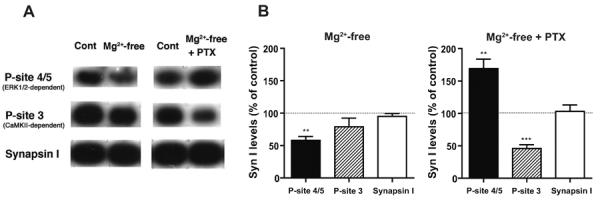
A, Representative immunoblots showing the phospho-site 4/5 (P-site 4/5), phospho-site 3 (P-site 3) and total levels of synapsin I in cortical slices incubated in different conditions. The ERK1/2-dependent-phospho-site 4/5 level was decreased in slices incubated in Mg2+-free condition alone, while it showed a marked increase in slices treated with concurrent GABAA-R blockade by picrotoxin (PTX, 100 μM), compared to control slices incubated in parallel in normal ACSF (Cont). In contrast, the phospho-site 3 level that is dependent on CaMKII, but not on ERK1/2, was unaltered in Mg2+-free condition alone, and largely decreased in Mg2+-free condition with picrotoxin. The total synapsin I level remained unchanged in either condition. B, Quantitative data obtained by immunoblot analyses, expressed as a percentage of control levels. ** p<0.01, *** p<0.001, one group t-test, n=5.