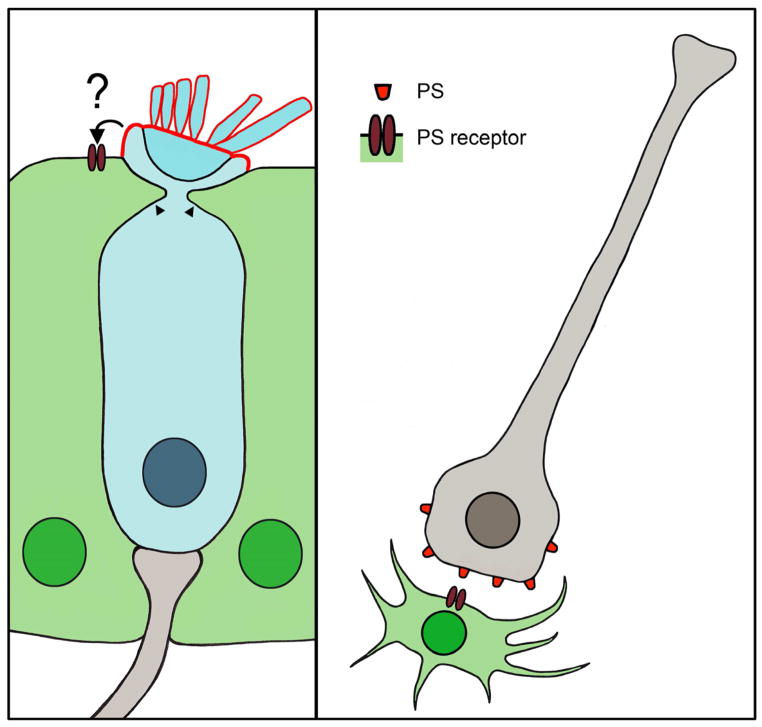
Figure 2.
Supporting cells and glia eliminate dead cells. Left panel, illustration of supporting cell processes (arrowheads) invading a neighboring hair cell (blue) during the process of hair bundle excision. Phosphatidylserine (PS, red outline) exposure is restricted the apical membrane of the hair cell, but its interaction with a PS receptor on neighboring supporting cells is unclear. Right panel, illustration of a neuron (gray) as a microglia (green) approaches for phagocytosis. The damaged neuron exposes PS, which binds a PS receptor expressed by the microglia.

