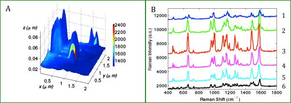
Figure 1.

(A) TERS map with simultaneously obtained topographic features and Raman characteristics from 80nm biotinylated-GNP probe adsorbed onto a streptavidin functionalized slide. Scan size: 2×2 μm, TERS step-size 55nm. Color represents the Raman intensity of a streptavidin marker band (965cm−1). (B) Plot of the representative TERS spectra extracted from 6 pixels along the colored line y= 1.08 μm in (A), where (1) x=1.31 μm, (2) x=1.25 μm, (3) x=1.14 μm, (4) x=1.08 μm, (5) x=0.97 μm, (6) x=0.86 μm.
