
Figure 8.
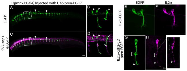
A – D) Tg(mnx1:Gal4-VP16) embryos were injected at the 1-cell stage with a plasmid encoding pren-EGFP under a 14X-UAS element, fixed at 24 hpf, and immunostained with SV2 and znp1 antibodies. Arrowheads point to primary motor neurons expressing EGFP. B, D) higher magnification images of the squares drawn in A and C respectively. Arrowheads point to primary motor neurons somas. E – I) Tg(mnx1:Gal4-VP16) embryos were injected at the 1-cell stage with the 14X-UAS-IL2α-EGFP plasmid (E, F) or with 14X-UAS-IL2α-CD & pren-EGFP plasmid (G – I). Embryos were fixed at 24 hpf, and immunostained with anti-IL2α antibodies (F, H, and I). IL2α-EGFP is evenly distributed throughout the motor neuron (E, F). Pren-EGFP labels the entire cell body and axon (G) while IL2α-CD is detected as discrete puncta through out the cell (H, arrowheads). I) Higher magnification of the axon shown in (G) indicating IL2α-CD puncta accumulated at the choice point (bracket) and at the distal tip of the axon. Scale bar in C, 50 μm; Scale bar in D, 10 μm; Scale bar in F and H, 10 μm. Dorsal is to the top and rostral is to the left.