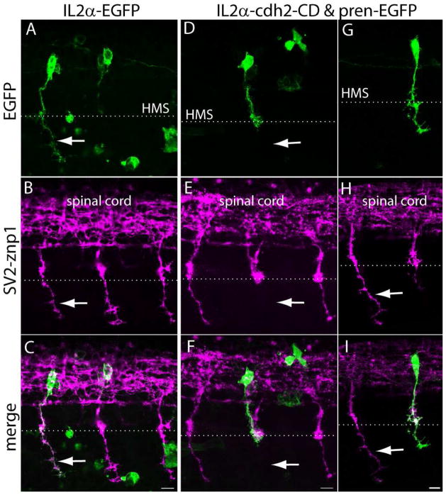
Figure 9.
Expression of N-cadherin dominant-interfering cytoplasmic domain perturbs CaP motor axon growth at the horizontal myoseptum. A – I) Tg(mnx1:Gal4-VP16) embryos were injected at the 1-cell stage with IL2α-EGFP (A – C) or with IL2α-cdh2-CD & pren-EGFP (D – I) plasmids. Embryos were fixed at 24 hpf, immunostained with SV2 and znp1 and observed under confocal microscopy. A – C) The arrow points to the CaP axon extending ventral to the horizontal myoseptum (HMS) indicated with a dashed line. The IL2α-EGFP labeled axon has a normal morphology as compared with the untransfected neighboring axons labeled with SV2 and znp1. D – F) A motor axon expressing IL2α-cdh2-CD grew through the common pathway but stalled at the horizontal myoseptum. The arrow points to the absence of SV2 and znp1 labeled CaP axon ventral to the choice point while neighboring untransfected axons grew normally. G – I) A CaP motor neuron expressing IL2α-cdh2-CD and pren-EGFP in which the axon extended ventrally to the horizontal myoseptum but shows a shorter migration distance as compared with an untransfected SV2 and znp1 labeled axon (arrow). Scale bar in C, F, and I, 10 μm. Dorsal is to the top and rostral is to the left.

