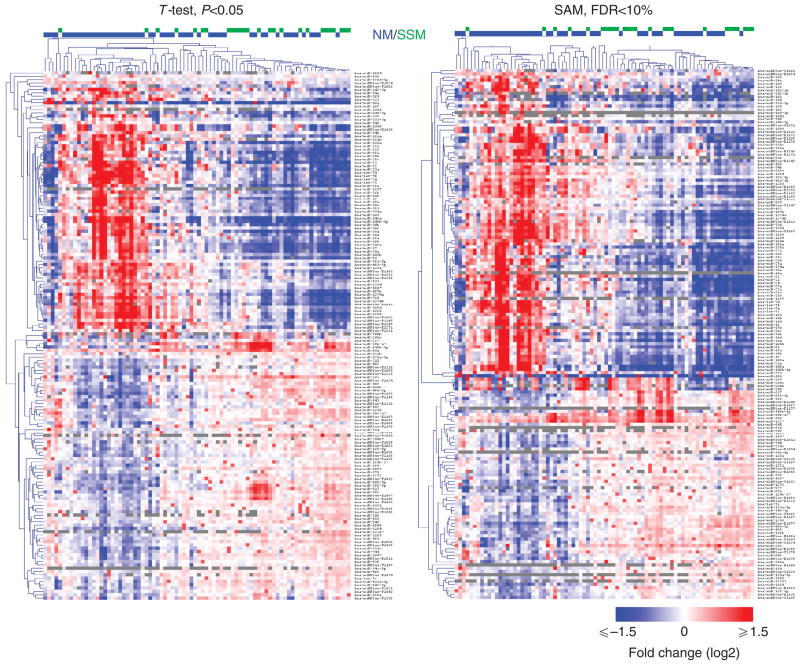
Figure 1. Expression profiling reveals 134 microRNAs that are differentially expressed between superficial spreading melanoma (SSM) and nodular melanoma (NM).
Heat map representing the differentially expressed microRNAs according to T-test, P<0.05 (left), and SAM, false discovery rate (FDR) <10% (right). T-test and SAM identify 160 and 165 differentially expressed microRNAs, respectively. In all, 134 microRNAs overlap between the two analyses. For 126 of the 134 microRNAs, the differences between SSM and NM could not be accounted for by thickness.

