
Figure 3. Transiently enhanced pulmonary infiltration of CD4+ and CD8+ activated T cells is independent of TNF produced by myeloid cells during M. tuberculosis infection.
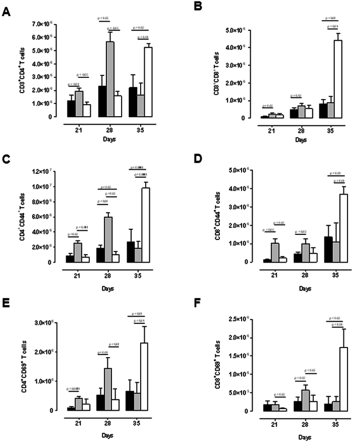
WT (black), M-TNF−/− (grey) and TNF−/− (clear) mice were infected via aerosol inhalation with 200–500 cfu/lung of Mycobacterium tuberculosis. Lung cell suspensions from infected mice were analyzed by flow cytometry to determine (A) CD3+CD4+ and (B) CD3+CD8+ T cell pulmonary populations at day 21, 28 and 35 post-infection and activation status analysed for markers CD44 (C and D) and CD69 (E and F) on CD4+ (C, E) and CD8+ (D, F) T cells by flow cytometry. The results are expressed as the mean ± SD of 5 mice per group and are from one experiment, representative of two independent experiments.