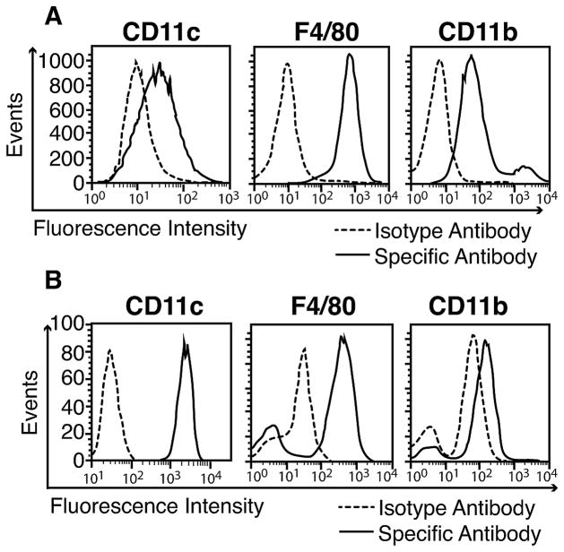
Figure 4.
Phenotyping of mouse alveolar macrophages. A, MH-S macrophages were stained with anti-CD11c, anti-F4/80 and anti-CD11b antibodies while control cells were exposed to fluorochrome labeled isotype antibodies. Cells were then analyzed by flow cytometry. B, Primary alveolar macrophages were obtained from normal C57BL/6 mice by broncho-alveolar lavage. The abundancy of CD11c, F4/80 and CD11b surface markers was detected by flow cytometry as described above. Data are representative of 2 independent experiments.

