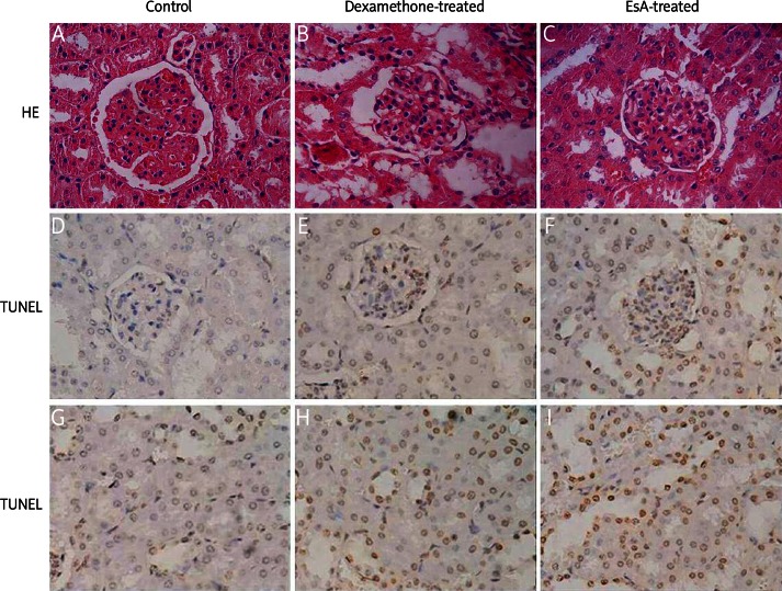
Figure 1.
HE staining showed that the mesangial cell proliferation and mesangial matrix decreased and capillary opened in the dexamethasone-treated group and EsA-treated group (A-C). TUNEL positive cells (yellow particles in the nucleus) were apparent in glomerular cells and tubular epithelial cells (D-I). Magnification: 400×

