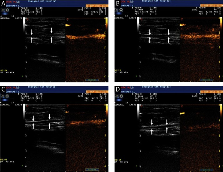
Figure 5.
Contrast-enhanced imaging of the carotid artery by the intravenous injection of cationic liposomal microbubbles (arrow indicates the carotid artery): A – images of the carotid artery at 4 s after CLM injection; B – images of the carotid artery at 25 s after CLM injection; C – images of the carotid artery at 123 s after CLM injection; D – images of the carotid artery at 174 s after CLM injection
CLM – cationic liposomal microbubble

