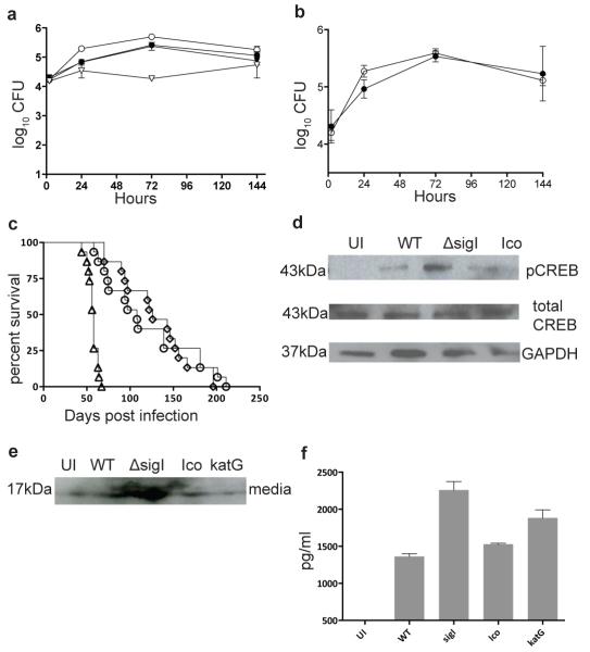
Figure 4. M.tb. ΔsigI mutant strain is not attenuated in cells or mice.
(a) J774A.1 murine macrophages were infected with wild-type M.tb. (closed circle), the ΔsigI mutant (open circle), the complemented strain (closed triangle), and the ΔkatG mutant (open triangle). (b) J774A.1 cells were co-infected with wild-type M.tb. (closed circle) and the ΔsigI mutant (open circle). (c) Time-do-death for three groups each of 20 Balb/c mice that were infected with an inoculum that implanted ~ 3.7 log10 bacteria per lung by aerosol; wild type (open circle), sigI (open triangle), and complement strain (open diamond). (d) CREB phosphorylation was measured by Western blot in M.tb.-infected J774A.1 cells at 12h. (e) TNF-α production was measured in infected J774A.1 by Western blot. (e) TNF-α production was measured in infected J774A.1 by ELISA. The time-to-death experiment was performed once. Three biological replicates of all pther experiments were performed, and error bars represent standard deviation.

