Fig. 6.
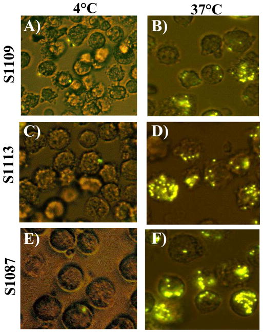
Internalization of phagocytosed beads by hamster lymphoma cell lines measured by confocal microscopy. Cells are shown 120 min after the addition of 30 fluorescent-labeled beads per cell and incubation at either 4°C (Panels A, C, E) or 37 °C (Panels B, D, F). Shown are hamster cell lines S1109 (Panels A, B), S1113 (Panels C, D), and S1087 (Panels E, F). Original magnification, 20×.

