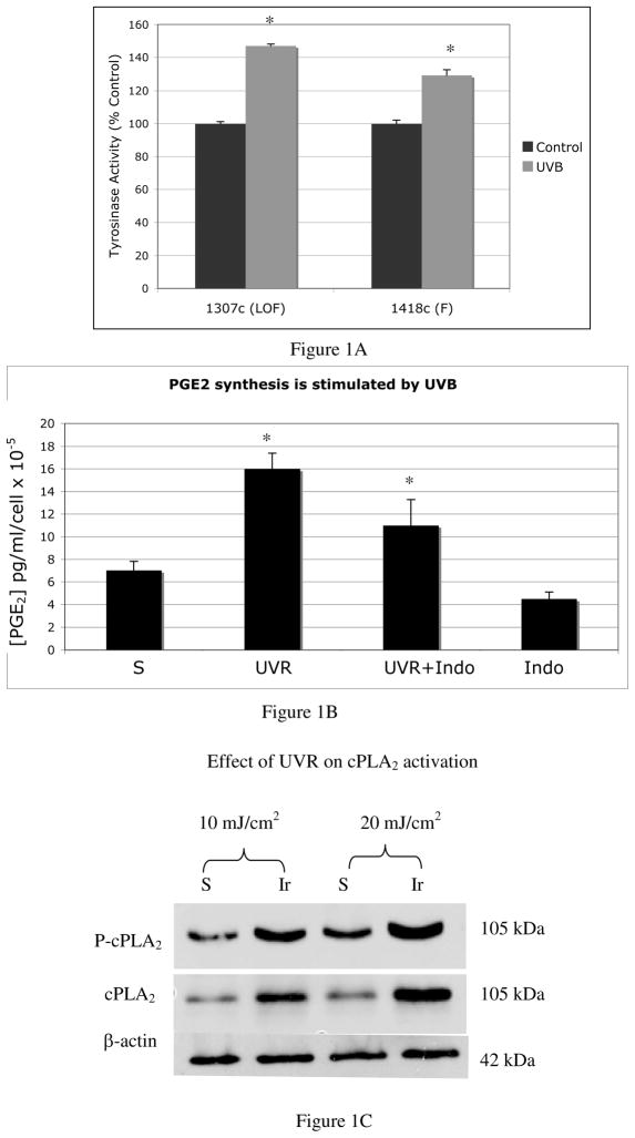
Figure 1. Evidence that the UVR-induced autocrine factor is not α-MSH, and that UVR stimulates PGE2 synthesis.
A). Tyrosinase activity (represented as percent of controls) is increased in loss-of-function (LOF:1307c) MC1R mutants by UVR (*p<0.005), indicating the presence of an autocrine factor that is not α-MSH stimulates tyrosinase activity in response to UVR. Results are the average of four separate experiments performed on melanocytes cultured from MC1R mutant melanocytes (1307c) and melanocytes with confirmed functional MC1R (1408c) +/− standard error of the mean (SEM).
B) Melanocytes showed a 2-fold increase in levels of PGE2 in response to UVR, which was inhibited by indomethacin, indicating that COX enzymes are activated by UVR in melanocytes. Differences in PGE2 were statistically significant between irradiated and sham irradiated cells, and between irradiated cells and cells irradiated in the presence of indomethacin (*p=0.12). Each bar represents the average amount of PGE2 from 3 independent experiments, +/− SEM, in which each condition was performed in triplicate wells, and in which each experiment was performed using melanocytes cultured from a separate Caucasian donor (n=3).
C) UVR induced the phosphorylation of cPLA2 and increased levels of cPLA2 protein in melanocytes. Results are representative of 2 experiments. For each experiment, melanocytes cultured from a separate Caucasian donor were used (n=2).

