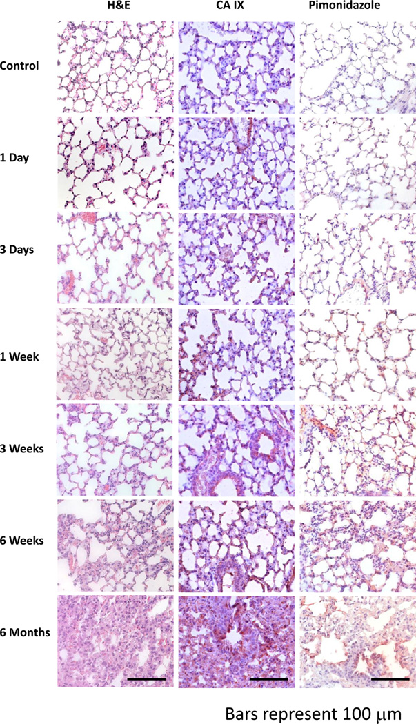
Figure 1. The temporal progression of tissue hypoxia in lung after radiation.
Tissue hypoxia is first noticeable around three days (CAIX, Pimonidazole) post-radiation and progressively increases throughout the follow-up period (6 months). At six weeks, the first histopathologic lesions are seen (H&E). These are generally focal in nature and are characterized by thickening of the alveoli wall and increased inflammatory cell infiltrate. By six months, tissue damage had considerably worsened and a greater number of focal lesions were observed. Error bars represent 100 µm.

