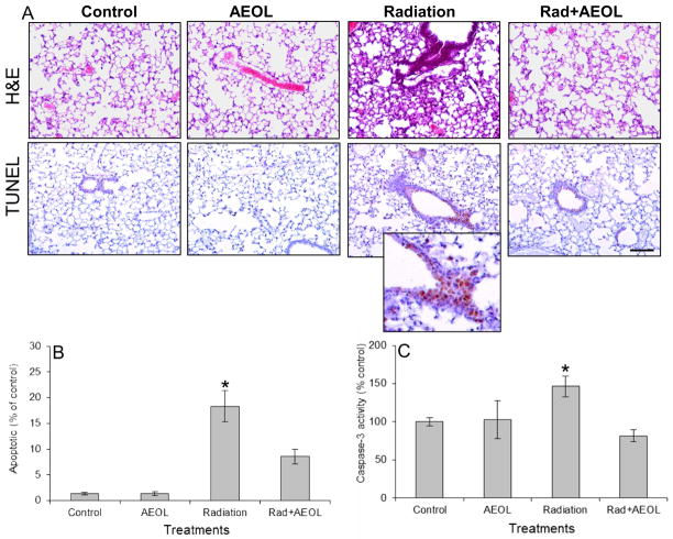
Figure 1. Apoptosis in lung tissue six weeks after thoracic irradiation.
(A) The increase in apoptotic nuclei (TUNEL assay) in irradiated lung tissue is reduced in AEOL10150 + IR treated mice. (B) Apoptotic index indicates the average number of apoptotic nuclei per 20x field normalized to sham-irradiated controls. An increase in positive staining for apoptotic nuclei was observed in irradiated lung tissue six weeks post-exposure which was not observed in mice treated with AEOL10150 for four weeks after exposure. Bar represents 100 μm. *p < 0.001 Rad vs. control; *p < 0.05 Rad vs. Rad+AEOL group. (C) Normalized caspase-3 activity confirms the presence of apoptosis in irradiated tissue. *p < 0.05 Radiation vs. each of the other groups.

