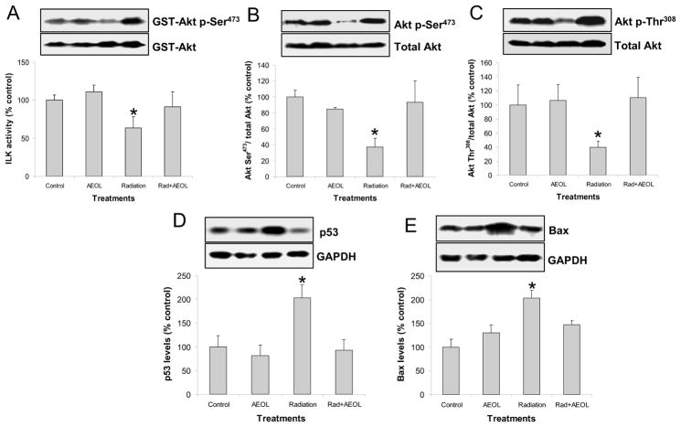
Figure 4. Changes in PTEN signaling.
(A) ILK activity assay in lung tissue lysate (*p < 0.05 vs. each of other groups). Top panel shows representative Western blot of phosphorylated GST-Akt (activated by ILK) and unphosphorylated GST-Akt substrate. (B) Significant decrease in Akt phosphorylation at Ser473 (*p < 0.05) and (C) Thr308 residues were observed in irradiated lung. (D) p53 (*p < 0.05) and (E) Bax (*p < 0.01 vs. control group; *p < 0.05 vs. AEOL alone and RAD+AEOL groups) protein levels were significantly increased in irradiated lung tissue 6 weeks after radiation. The top panels in D and E show representative Western blots of p53 or Bax and GAPDH.

