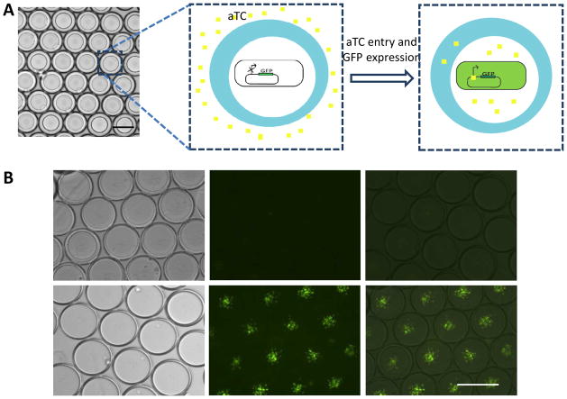
Figure 3.
aTC diffusion and activation of GFP expression. A) Schematic illustration of the experiment design. GFP was expressed under aTC dependent promoter. Bacteria carry this expressing vector was encapsulated in the core without aTC. aTc was then added to the external medium, which diffuse into core of droplets to activate GFP expression; B) Fluorescence microscope images of droplets containing bacteria without aTC addition (top panel) and with aTC addition (bottom panel). Images were taken 1 hour after cell encapsulation. (Scale bar: 100 μm)

