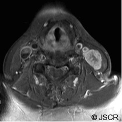
Figure 1.

MR and CT Neck findings of the surgical pathway recurrence of a clival chordoma. (Axial, T1-weighted with Gadolinium MR image). Image obtained at the time of presentation with the neck mass, show a large, heterogeneous level III neck mass (23 mm x 17mm) with infiltration throughout the sternocleidomastoid muscle without obvious invasion of the great vessels. A few surrounding rounded lymph nodes within normal size limits are seen.
