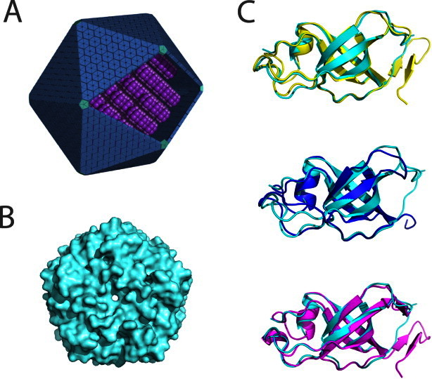
Figure 1.

Comparison of the structure of GrpN with other bacterial microcompartment vertex (BMV) proteins. (A) An idealized model of an MCP showing pentameric units at the vertices of the polyhedral shell. (B) Space-filling model of the pentameric structure of GrpN. (C) Superposition of a GrpN monomer with CcmL (top, yellow), CsoS4A (middle, blue), and EutN (bottom, magenta). [Color figure can be viewed in the online issue, which is available at wileyonlinelibrary.com.]
