
Figure 2.
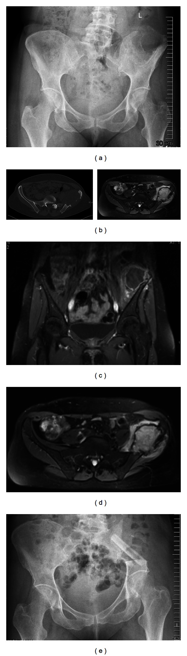
(a) Anteroposterior radiograph of the pelvis, showing a large osteolytic lesion of the left iliac bone (synovial sarcoma). (b) CT scan of the same patient showing the size of the tumor. Notably is the lack of matrix or calcification inside the tumor. (c) and (d) MRI scan of the same patient showing the intra- and extrapelvine size. (e) Postoperative X-ray after P1 resection and pelvic reconstruction stabilised with an autologous nonvascularised fibular graft.