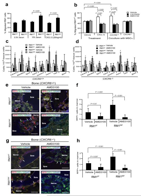
Figure 5. EMT-mediated CXCR4 is highly involved in prostate cancer metastasis.
(a) Migration assays were performed in Transwell® plates using 10% serum or CXCL12 as chemoattractants. Migration toward 0.5% serum was used as a negative control. (b) Blockade of CXCR4 by AMD3100 or anti-CXCR4 antibody prevents prostate cancer migration towards CXCL12 or MSCs isolated from CXCR6+/+, but not CXCR6−/− animals. Data in (a,b) are representativedata from two independent studies (mean±s.d., ANOVA). Significance was determined using a Student’s t-test. RFP-labeled RM1WT or RM1EMT cells (Supplementary Fig. S5a) were incubated with vehicle or AMD3100 in vitro, and then inoculated by intra-cardiac (i.c.) injection into CXCR6+/+ or CXCR6−/− (n = 7). Metastasis was assessed by qPCR for RFP in a number of tissues. (c,d) Number of metastatic RM1 cells following i.c. injection. *Significance between RM1WT treated with vehicle and RM1WT treated with AMD3100 (P < 0.05). #Significance between RM1WT treated with vehicle and RM1EMT cells treated with vehicle (P < 0.05). †Significance between RM1EMT treated with vehicle and RM1EMT treated with AMD3100 (P < 0.05). Error bars represents mean±s.d., n = 2 independent experiments, P < 0.05; Student’s t-test. (e-h) RM1 cells expressing RFP were identified in the femur of CXCR6+/+ or CXCR6−/− mice following i.c. injection. Red arrows identify RM1 cells. White arrows identify osteoblast on the bone surface staining positive for CXCL12 expression. Scale bars, 100μm. (f,h) Quantification of Fig. 5e and Fig. 5g, respectively. The numbers of RM1 cells were quantified on the endosteal region of the 7 long bones. Endosteal regions were defined as 12 cell diameters from bone surfaces. ((Mean±s.d. (n = 3)., ANOVA).

