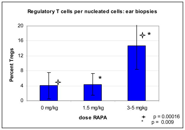
Figure 5. Percent FoxP3 positive cells per total infiltrating nucleated cells.
Immunohistochemistry staining was done on the ear biopsies from day 30 for FoxP3. A percentage of FoxP3 positive cells per total nucleated infiltrating cells were analyzed. 1000 cells, or maximum possible cells were counted to determine percentage. Above graph represents slides obtained in three separate experiments, in 0 mg/kg group n = 10; in 1.5 mg/kg group, n=4; in 3–5 mg/kg group, n=11.

