
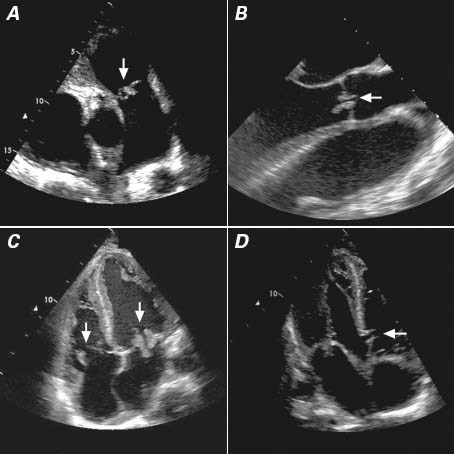
Fig. 1 Transthoracic (A, C, D) and transesophageal (B) echocardiographic images show vegetations (arrows) on the A) pulmonary valve, B) aortic valve, and C) tricuspid (left) and mitral (right) valves, together with D) a perimembranous ventricular septal defect (arrow).