
Figure 1.
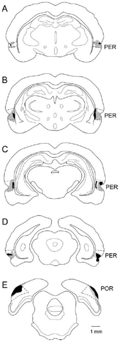
Combined perirhinal (PER) and postrhinal (POR) lesions. Schematics of coronal sections through the rat brain are shown with the largest lesion in gray and the smallest in black. Lesions were relatively small but distributed along the rostrocaudal extent of the PER and POR.