
Figure 1.
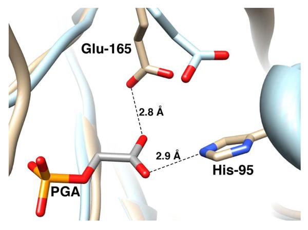
Models, from X ray crystal structures, of the active site of unliganded yeast TIM (light blue, PDB entry 1YPI) and yeast TIM complexed with PGA (tan, PDB entry 2YPI). Ligand binding is accompanied by a 2 Å shift in the position of the carboxylate side chain of Glu-165 towards the bound ligand. The active sites of TIM from yeast and Trypanosoma brucei brucei are virtually superimposable, except that the catalytic base at TbbTIM is at position 167 in the protein sequence.