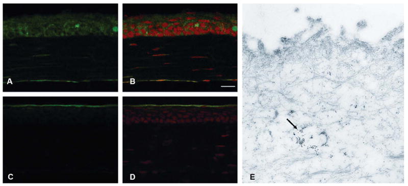
FIGURE 1.

Constitutive immunolocalization of nonphosphorylated HSP27 in unwounded corneas (A) and merged image with PI staining (B). Localization of phosphorylated HSP27 in unwounded corneas (C) and merged image with propidium iodide staining (D). Localization of phosphorylated HSP27 in unwounded corneas by immunogold electron microscopy (E, indicated with black arrow). Nonphosphorylated HSP27 was present in all epithelial layers of unwounded mouse corneas (A and B), whereas phosphorylated HSP27 was localized only in the superficial epithelium of unwounded corneas (C–E).
