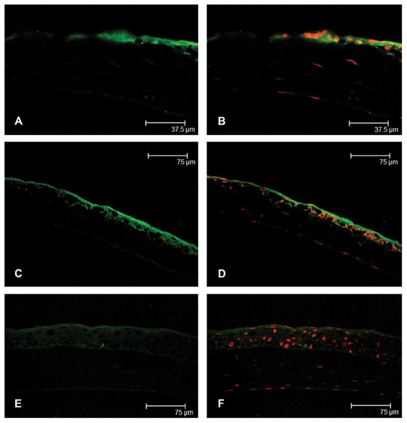
FIGURE 3.

Immunolocalization of phosphorylated HSP27 from the wounded central cornea (A and C) to the unwounded peripheral cornea (E) at 8 hours after epithelial debridement and merged images with propidium iodide staining (B, D, and F), respectively. The epithelialization was not complete, and phosphorylated HSP27 expression was prominent at the leading edge of the healing central corneal epithelium (A and B). Phosphorylated HSP27 in wounded central cornea was localized to the superficial and basal epithelial layers, and the staining intensity was similar in all epithelial layers (C and D). However, phosphorylated HSP27 in unwounded peripheral corneal was localized only to the superficial epithelium (E and F).
