
FIGURE 4.
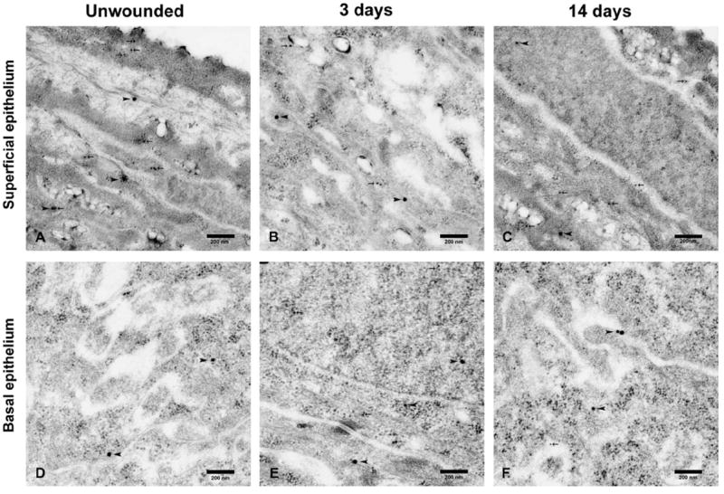
Detection of nonphosphorylated HSP27, indicated by arrow head, and phosphorylated HSP27, marked by arrow, by double immunogold labeling transmission electron microscopy in unwounded superficial (A) and basal (D) epithelial layers, superficial epithelial layers of wounded corneas after 3 days (B) and 14 days (C), and basal epithelial layers of wounded corneas after 3 days (E) and 14 days (F). The distribution of nonphosphorylated HSP27 remained unchanged in the superficial and basal epithelium over 3 time points. The distribution of phosphorylated HSP27 in the basal epithelium was sparse in unwounded corneas (D) and increased after 3 days (E) and was sparse again after 14 days (F), whereas that in the superficial epithelium did not differ as time went (A–C).