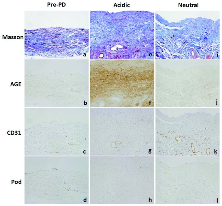
Figure 1.
— Peritoneal histology and immunohistochemistry for advanced glycosylation end-products (AGEs), CD31, and podoplanin (Pod) in (a-d) the pre-peritoneal dialysis (PD) group and (e-h) the acidic and (i-l) neutral dialysate groups. 200× original magnification. Peritoneal samples from the acidic dialysate group show significant fibrosis in the submesothelial compact zone, with accompanying hyalinizing degeneration of collagen fibers. In the acidic group, AGEs also accumulated more intensely. Staining for CD31 revealed increased vascularity in the neutral group; however, staining for podoplanin revealed no increase of podoplanin-positive lymphatic vessels, and no differences between the groups. Masson = Masson trichrome stain.

